Taking Control of Your Concussion Recovery

Check out my February post and other great advice at Concussion Connection.
Key excerpt:
“In certain situations, less might be more by building in easier days, rest days, or even off weeks.”
Please Check Our New Brand and Website: www.ActiveKidMD.com
Comprehensive blend of general pediatric and sport medicine care with an individualized approach that enhances the health and knowledge of patients and their families
ACCEPTING NEW PATIENTS- CALL 714-974-2220 FOR AN APPOINTMENT
CLICK HERE FOR DR. KOUTURES GENERAL PEDIATRICS INFORMATION
Proud physician:
USA Volleyball Mens/Womens National Teams
CS Fullerton Intercollegiate Athletics
Chapman University Dance Department
Orange Lutheran High School
Co-Author of Acclaimed Textbook
Pediatric Sports Medicine: Essentials for Office Evaluation
Orange County Physician Of Excellence, 2015 and 2016
Check out my February post and other great advice at Concussion Connection.
Key excerpt:
“In certain situations, less might be more by building in easier days, rest days, or even off weeks.”
1. In youth soccer, most lower body injuries come from non-body contact and occur more in competition than training or practice sessions. While training injury incidence rates usually do not change with increased player age, match injury incidence tends to increase with age through all age groups
2. The time of the adolescent growth spurt (girls usually age 12-14 and boys usually age 13-16) seems to have an increased vulnerability for traumatic injuries. Afterwards athletes seem to be susceptible to cumulative overuse injuries.
3. Knee injuries occur in 7% to 36% of injured players and are seen more frequently in females Middle school soccer playing females have a higher rate of anterior knee pain issues than volleyball or basketball players. Any single-sport adolescent female has a higher risk of anterior knee pain issues.
4. Adolescent female soccer players suffer a roughly 3-6 times increased risk of ACL rupture compared to boys playing the same sport. Several factors have been proposed for the increased risk, such as anatomic differences, hormonal contributions with menstrual cycles, and higher-risk single-leg landing, turning, and jumping positions.
6. Ankle injuries account for 16% to 29% of injuries and are more frequent in male and older players Ankle contusions more common in younger players due to the more ground-oriented game, while in older players ankle sprain tend to occur due to the more aggressive and faster game.
9. Greater exposure to training and competition leads to a greater risk of injury due to the high intensity of the activities.
What ideas do I have to help reduce these risks?
What ideas would you add to help young soccer players reduce lower body injuries?
Do you know your child's age in years?
Can you remember the number "2"?
Good.
Those basic pieces of information allow you to make key decisions that can reduce the risk of overuse injuries in your young athlete.
If the number of hours of organized sport activity per week exceed the number of years of the age of a young athlete, then there is a statistically higher chance of suffering a serious overuse injury.
If the ratio of organized sports to free play is greater than 2:1, then there is a statistically higher chance of suffering a serious overuse injury.
That's it.
Pretty simple. Pretty easy to remember. Pretty easy to put into practice.
Thanks to Sports-Specialized Intensive Training and the Risk of Injury in Young Athletes: A Clinical Case-Control Study by trusted colleagues Neeru Jayanthi, Cynthia LaBella and their co-authors in Chicago, these simple decision rules can now provide evidence-based guidance to an area where concrete recommendations were sorely lacking. Over 800 injured 7-18 year-old athletes who were treated at two sports medicine clinics were compared to similar aged healthy children who came to the same clinics for pre-participation sports physicals.
Now, what are organized sports?
Any sport activity which is organized and supervised by an adult.
This does include games, practices, conditioning, speed training, weight training, and individual skills training sessions. Probably fair to extrapolate to technique courses, choreography courses, rehearsals, and individual skills sessions for dancers and other performers.
Not only do we get those two helpful decision rules from these findings, but also an emphatic reminder of the value of free play in the safe development of young athletes.
That's another simple thing to remember and put into regular practice.
Sincere thanks to Matt Swift, DPT from Change Sports Physical Therapy Institute for creating this video on Stress Injuries to the Tibia with focus on symptoms, treatment, and prevention.
Is it OK to exercise when sick? What symptoms should keep someone off the playing field?
When trying to decide if an athlete is too ill to participate in sports, I tend to ask the following questions:
Study and experience tells us that a fever over 100.4 degrees Fahrenheit may increase metabolic demands of the body, often making exercise more difficult. Thus, many authorities recommend starting with lighter levels of exercise with a fever, and using overall performance to advancing to higher intensity of exercise. Some athletes may perform quite adequately with a fever, while others will need complete rest from exercise until the fever is gone for at least 24 hours.
I have found that the neck rule can also assist athletes and parents in deciding on sport participation, with or without a fever:
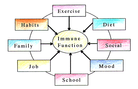
How can we limit spread of colds and other illnesses during the winter months?
Multiple factors contribute to healthy function of the immune system in preventing illness
No athlete wants to be sick during the season and no athlete wants to spread illness to other team members. Following some basic rules can help prevent the spread of infections:
How does the amount of exercise influence risk of getting sick?
Moreira A et al. Br Med Bull 2009;90:111-131, © The Author 2009. Published by Oxford University Press. All rights reserved. For permissions, please e-mail: journals.permissions@oxfordjournals.org
The above figure demonstrates that both intensity of exercise and fitness level can influence the risk of upper respiratory infection. More moderate levels of exercise can be protective against illness, while lower levels and higher levels of exercise may actually be related to an increased risk. This is often why athletes get sick right after higher level competitions or at the end of a particular sport season. Higher levels of fitness may also be protective against respiratory illnesses.
There is no magic age at which a child can begin weight training. Readiness for weight training depends on the willingness of the child to lift weights, follow directions, and maintain the program for several months to see results. Remember, this is for the child, not for an adult or coach.
Weight training should supplement regular sport activity. It is not acceptable to have weight training injuries keep an athlete away from his/her sport. I recommend qualified supervision by a performance or physical trainer who routinely works with children and adolescents. The focus should be on appropriate-sized equipment, meticulous weight lifting technique, starting with low weights/high repetitions, and working multiple body parts. In appropriate program, a child will often lift weights 2 or 3 days a week with at least 48 hours of rest between sessions.
The physical results, such as muscle enlargement and weight gain, depend on the gender and developmental stage of the child. Routine weight training can make a child somewhat stronger by increasing nerve and muscle communication. However, if the child is looking for larger and more bulked muscles, then they must wait until after their growth spurt. Androgens are a particular hormone, produced more in boys than girls, which produce muscle and strength gains. Since androgens increase late in puberty right after the growth spurt (age 11-12 in girls, age 13-14 in boys) lifting before this time will not result in massive muscle bulking or extreme strength gains.
Does this mean one should not lift before the growth spurt? No, but just place the emphasis on good technique and reduce the expectations for big-time muscle gain. Remember, due to lower androgen production than boys, girls will have less increase in muscle mass.
Is weight training safe for children?
Studies have shown that a properly designed and supervised resistance training program can be safe for children and young adults. Contrary to popular belief, weight training at a young age does not stunt growth as long as proper techniques are utilized. There are reports of overuse injuries with back strains the most common but at no greater frequency than what is seen on the athletic field. Again, placing the emphasis on a properly designed and supervised resistance training program will help reduce injuries and maximize enjoyment.
Click here to learn about:Proper post-lifting recovery, focusing on nutrition and sleep, can greatly enhance the results and safety of a weight training program.
Does weight training work?
Both published studies and personal experience have shown impressive strength, speed, and endurance gains with an appropriate weight training program. There is no good scientific data to show that this directly translates to better on-field performance, but it does contribute to overall athletic ability. The athlete needs to be aware that he/she must stay with the program or risk losing the gains.
To produce optimal results, recommend starting a program during break periods between sport seasons and not initially scheduling weight training sessions on same days as practices or games. Once the athlete is more comfortable with the demands of weight training, can incorporate lifting sessions with regular training and competition activities.
Can weight training reduce injuries?
High school-based studies indicate a resistance training program could decrease the number and severity of injuries, and also reduce the rehabilitation time once an injury has occurred. These benefits may be due to stronger supporting joint structures, muscle absorbing more energy before tiring out, and greater muscle balance around a specific joint.
Can weight training help with weight loss or weight control in children?
Weight training programs that feature higher repetitions, lower weights and limited rest between sets have been shown to contribute to both weight loss and weight control in children. Appropriate professional supervision in designing such a program can be of significant help.
For centuries, athletes have searched for any substance or technique that could enhance exercise and allow for more effective weight gain/loss or increases in strength and endurance. Many available performance enhancing products may report claims of potential amazing efficacy but use of them can be clouded by concerns over true scientific support, side effects, and financial cost.
Here are 5 sensible tips to guide you on nutrition and recovery with your exercise program:
1. The Importance of Sleep
Can’t begin to tell you how sleep deprivation can derail even the best constructed exercise program, as skeletal muscle needs adequate recovery time to rebuild damaged fibers and to increase the working capabilities of contractile units. Multiple studies support the efficacy of a minimum of 8-9 hours of sleep a day to foster such recovery. Insufficient sleep can also reduce mental alertness on the job or at school and has also been associated with statistically higher risk of illness or injury. Establish a regular bedtime and not allow deviation of more than ½ hour and also encourage daytime naps of under one hour per day which have been shown to be restorative, add to the cumulative daily sleep amount, and not adversely affect nighttime sleep patterns.
· TIP TO ASSIST WITH SLEEP: Stop any type of screen device use no later than one hour before bedtime, and do not have screen devices in the sleep area, as use right before bedtime or alerts/temptation to check during sleep have been associated with reduced amount and quality of sleep.
2. The Timing and Amount of Protein
Protein is the building block of skeletal muscle and is needed to assist in that reparative and rebuilding process after exercise. Good data suggests that the best time for workout-related protein intake is within 30 minutes after completing exercise. A post workout protein amount of 25-30 grams along with a total daily intake of 0.5-0.7 mg protein/pound of body weight are both solid recommendations. I have always favored dairy or meat/bean/egg sources of protein as readily available products that confer well-absorbed collateral benefits of calcium, Vitamin D, and iron. Amino Acids are the building blocks of proteins, and intake of specific individual amino acids has been touted for both weight loss and strength building. However, there is a lack of rigorous support for high amounts of individual amino acids, so stick with whole food protein sources.
· TIP TO ASSIST WITH PROTEIN INTAKE: 8-12 ounces of chocolate milk within 30 minutes of exercise is a sensible recovery drink that has a ratio of carbohydrate to protein that allows enhanced transport of protein to recovering muscles. Other good post workout food-based protein sources include Greek yogurt and peanut butter.
3. What About Creatine?
Creatine is a natural substance that assists with regeneration of short-acting energy sources that fuel contraction in working skeletal muscle. Increased amounts of creatine in working muscles can potentially contribute to more intense workouts and also assist in muscle recovery after workouts. Powdered and liquid creatine supplement products have been studied, with trials that include higher loading doses for the first few days, followed by lower daily maintenance doses and other regimens that include medium daily doses without any higher loading amount. Positive results have included increased speed and ability to complete multiple short-burst activities such as 80-100 yard sprints. Possible side-effects include water retention, bloating, muscle cramping, and potential kidney injury (currently only reported with individuals that had pre-existing kidney concerns). Anecdotally, many athletes using creatine have reported enhanced recovery with increased ability to work harder in subsequent workouts, and documented strength increases support increased muscle size not being simply due to water retention, but to increased contractile abilities. Take note that many United States-based sports medicine advisory organizations do not endorse creatine supplementation in children under 18 years of age.
· TIPS ON CREATINE USE: Creatine monohydrate in either liquid or powder format has been the best studied form of creatine. If using any supplement, ensure that you are getting exactly what is on the container and not any additional substances (see below). Reductions in creatine dose have been shown to assist with bloating, cramping or extreme water retention. Good food sources of creatine include wild game meats or wild-caught fish which also have those collateral benefits of protein, calcium and iron. Domestic meats and fish (especially free-range meats) still have reasonable amounts of creatine.
4. Are Pre-Workout Supplements Safe and Efficacious?
Advertised preworkout supplements or “energy drinks” report to enhance athletic performance and routinely contain multiple components such as caffeine and taurine. While a few small studies support the performance enhancement of stand-alone agents, published data on combination products is scant, inconclusive, or confusing. Safety concerns exist with use of products that have cross reactivity of multiple agents or larger than studied amounts of a certain product.
· TIPS ON PREWORKOUT SUPPLEMENTS: Read labels! If using any preworkout supplement in addition to usual daily caffeinated beverage of choice, you might be getting an enormous caffeine load with possible dangers that outweigh possible benefits to your workout.
5. What’s the Lowdown on other Supplements?
Use of a vast array of pre and post-workout supplement products has been touted to enhance athletic performance and exercise capabilities. Many of these products will work- no doubt about it, - however the gross majority of products do not have any rigorous practical scientific result to support clams and there is a tangible risk of adverse consequences from both known and potentially unreported elements in supplement products. What is on the label of many products is not the same as what is in the actual container. Personal experience along with results found by national sport organizations have found a significant number of supplements that have additional, unreported elements that are potentially dangerous and might be banned for sporting competition. While most of you are not subject to drug testing for performance-enhancing agents, suffering real and possibly long-term health consequences is not worth the short-term gains in strength and endurance.
Each concussion deserves individualized recommendations that seek to strike the delicate balance between a child's need for maintaining social contacts and attempt to continue with school work with a desire to not overwhelm the healing brain and increase post-concussion symptoms. An absolute restriction on screen use might reduce possibility of certain symptoms such as difficulty falling or staying asleep, but can also lead to social isolation contributing to higher symptom reports of anxiety, sadness, and outright depression.
How can we best strike an appropriate balance between screen use and need for adequate sleep?
Ask most parents if they have worries about sleep issues and amount of electronics/screen device use in their school aged children, and you'll probably get ready nods and smiles of affirmation.
Ask some of my sports medicine colleagues about why we are seeing more complicated and prolonged post-concussion recoveries, and you'll hear some suggest that the multi-tasking and multiple platforms of communication utilized by smart phones and other screen devices are potential contributing factors.
So since increasing sleep issues and attempts to pry screen-based devices from the hands of kids are common concerns to parents and medical professionals, it should be no surprise that difficulties initiating or maintaining sleep and regulating electronic use are often major challenges in children who have suffered a concussion.
Came across two recent studies on the subject of screen use and sleep that I think shed some interesting light on how we might make recommendations for all children, but particularly in the immediate post-concussion population.
One study from Proceedings of the National Academy of Sciences of the United States of America suggests the use of portable light-emitting devices immediately before bedtime has potential biological effects that may perpetuate sleep deficiency and disrupt circadian rhythms, both of which can have adverse impacts on performance, health, and safety. Such device use can:
While this study used healthy young adults (mean age around 25 years of age), the findings are intriguing enough to be extrapolated to younger patients. Given the frequency where recommended oral melatonin clearly helps with falling and staying asleep, having another pathway to support internal melatonin production can be essential in the recovery process.
An additional study from the journal Pediatrics examined 4th through 7th graders and assessed associations of different screens in sleep environments with sleep duration and perceived insufficient rest or sleep. Particular interest was placed on smartphones which can emit notifications during sleep periods, and relevant findings included:
These findings found that small screens could have more adverse effects on sleep than television screens and thus caution against unrestricted screen access in children’s bedrooms for normal, healthy 4th through 7th graders, which again could be extrapolated to include concussed children.
Throwing this all together, a pragmatic approach to screen use after concussion that utilizes the findings of these studies may include the following clinical recommendations:
1) The preponderance of screen devices is an integral reality in the life of many school-aged children and significance of appropriate use cannot be underestimated in expediting post-concussion recovery.
2) Once appropriate, limit screen device time use initially to the middle of the day and not within one hour of any scheduled nap or evening sleep period.
3) All screen device use should be stopped at least one hour before bedtime,
4) Screen devices should be removed from the bedroom to reduce interruptions in sleep from notifications or temptation to check devices for updates during periods of awakening.
Once the child has recovered from the concussion, the child might find that continuing the above screen time recommendations may lead to continued enhanced amount and quality of sleep, which in itself may lead to an enhanced quality of life.
As a sports medicine specialist, here 5 important factors that should be included in every evaluation of a bone stress injury:
1. Timing
2. Technique
3. Appropriate Energy Intake
4. Not enough rest
5. Growth spurts
This blog post does not intend to diagnose or provide any management tips for a particular stress injury, or any other injury or illness. If you suspect a stress injury, please immediately contact a sports medicine specialist for appropriate evaluation and treatment recommendations.
Overload injuries to bone are aptly called stress injuries as their often untimely presentation and unpredictable healing times can provoke high levels of emotional stress for patients and medical providers. While the actual diagnosis can require some detailed investigation, trying to identify root causes of stress injuries is a necessary detective game that can ultimately reduce the risk of future stress injuries and assess the overall bone health of the athlete.
Stress Fracture of the outer lining of tibia (shin bone) in a young dancer
Stress injuries to bones of athletes are often caused by relative overload, and in describing the spectrum of possible bone stress or overload injuries to patients, I often use the analogy of bending my pen while bored in class one day.
Stress Fracture of Left Femoral Neck: This MRI picture shows a fracture line involving only one cortex (outer lining) of the femur (thigh bone) with bone swelling (edema) also present.
Look forward to an upcoming post on important things to consider after the diagnosis of a bone stress injury has been made.
Early bird registration ends December 19, 2014. Register today!
We have exciting news about our upcoming Sports Cardiology & Sudden Cardiac Arrest in the Young on Friday-Saturday, January 23-24, 2015.
Keynote Speaker, Michael J. Ackerman, MD, PhD, from Mayo Clinic in Rochester, Minnesota will be participating several times throughout our 2-day conference and giving his keynote address: State of the Molecular Autopsy Following SCD on Saturday morning.
Dr. Koutures will speak on Heat Stroke: When Is It and When Is It Not? with emphasis on Prevention and Management of Exertional Heat Illness, Sickle Trait Collapse, Exertional Rhabdomyolysis and Exertional Hyponatremia.
One of the outcomes from this meeting will be to publish national guidelines on the "Prevention of Sudden Cardiac Arrest in the Young." A special panel discussion on Friday afternoon will directly impact these guidelines.
1) Osteochondral Lesions
Articular cartilage is thin tissue that covers the ends of the thigh bone (femur), shin bone (tibia) and backside of the kneecap (patella). An osteochondral lesion is damage to that creates crescent-shaped fragments (look like "shark-bites") of bone and cartilage that is most extreme cases may separate from the bone of origin and float within the knee joint. Symptoms may mimic more common anterior knee pain, though locking, catching, and local swelling are more suggestive of osteochondral damage.
Osteochondral lesions can be initially identified on plain x-rays, which must include the tunnel view which best visualizes the lower thigh bone. Have seen cases where not obtaining the tunnel leads to missed opportunities to identify injury, such at the osteochondral lesion identified on the inside of the femur seen on the x-ray image above. Magnetic Resonance Imaging (MRI) is often used to better characterize size and nature of lesions.
Management of osteochondral lesions depends on several factors:
2) Anterior Fat Pad Impingement
Image courtesy of http://www.physiotherapy.co.uk/blog/wp-content/uploads/2011/09/hoffas_impingement1.gif
Located below the patella and behind the patellar tendon between the femur and tibia (see yellow shaded region in adjacent picture), an enlarged or irritated anterior fat pad can become trapped and cause pain especially with bending of the knee. More commonly seen in adolescents, this is often best identified by direct finger-tip pressure placed on either side of the patellar tendon with the knee bent to about 90 degrees.
While identification of fat pad impingement can be a challenge, treatment is also fraught with unique potential challenges. Direct injection of anesthetic and anti-inflammatory medication can help both with diagnosis and pain relief, but often the initially promising results wear off within a few months. Surgical excision of the fat pad is a reasonable next option, but regrowth of the fat pad commonly can occur.
3) Placing Too Much Focus on the Knee
When the knee hurts, seems logical to put direct emphasis on correcting problems at that joint. However, failing to evaluate and respond to mechanical issues above and below the knee can slow progress and prolong pain and frustration.
This article is not designed to provide any diagnosis or treatment recommendations. Seek qualified pediatric sports medicine speciality evaluation to help young athletes properly identify factors causing chronic anterior knee pain and provide potential solutions.
I have accessed CoachSmart while on the sidelines, and no longer have to guess or try to remember suggested adjustments for practice and games in hot or humid weather. The information is concisely presented in the palm of my hand.
The iPhone app CoachSmart was developed by colleagues at Vanderbilt Sports Medicine and the Monroe Carell Jr. Children’s Hospital at Vanderbilt and is billed as the ultimate resource for coaches, offering real-time information on heat index and lightning strikes, frequently asked sports medicine and safety questions, and a group contact feature.
The The app is free to download in iTunes with an annual in-app subscription to live lightning data for $1.99.
Developed by Sports Medicine Physicians and Athletic Trainers with close guidance from coaches, the CoachSmart app brings many important topics into one easy location.
Recommending CoachSmart is now part of my pre-season safety talks to coaches, parents, and administrators, and will also be part of an upcoming lecture on Heat Illness.
The CoachSmart App was recently upgraded and returned to active status. I do not have any financial relationship with the CoachSmart App.
In an effort to better identify young athletes who might be a greater risk of prolonged recoveries after suffering a sport-related concussion, the findings of a recent retrospective study indicate that a personal or family history of mood disorders maybe linked to a longer recovery period.
Researchers at Vanderbilt University compared athletes who had a three week post-concussion symptom resolution period versus those with a three month or longer symptom recovery period, and found that those with pre-concussion anxiety or depression had a 17-fold increased risk of having the prolonged recovery time.
The research team also found that a family history of mood disorders and delayed onset of symptoms were both also associated with an increased risk of prolonged symptoms after a sport-related concussion.
These findings definitely mirror my experiences in working with school-aged athletes who have suffered concussions during sport activities.
I am more apt to ask about both personal and family history of mood disorders, often in the initial evaluation after a concussion. I will counsel families that any diagnosed or even suspected pre-existing emotional disorder, including Attention Deficit Disorder with/without Hyperactivity (not evaluated in the above study), depression and anxiety will have a tendency to worsen after concussion.
In some cases, it is the concussion that makes pre-injury issues more clear and out in the open, and in those instances, we both have to manage the concussion issues but also give proper respect and attention to those underlying mood disorders.
Early identification and aggressive psychological, medical, and school-based interventions are quite helpful in addressing emotional disorders. Key to have mental health colleagues available to assist in more difficult cases.
Failure to address the mood disorders leads to sub-optimal recovery.
I have also found that those pre-existing emotional disorders optimally managed with appropriate therapy and medication (when indicated) tend to have less consequences or flare-ups after an concussion.
The findings of delayed onset of symptoms is also not a major surprise. I do tend to see namely depressive symptoms not fully present for up to 4-6 weeks after a concussion. Not sure why this occurs. Could be part of the anticipated physiologic healing response, but could possibly be a by-product of cumulative mental and physical fatigue that accumulates by this time period and results in higher reported symptom presentations.
To read my post, please click here
For years, I have heard claims that some Major League Teams favor drafting pitchers who grew up in colder climates.
The reason?
Fewer months able to be spent outside likely means fewer competitive pitches thrown, fewer innings pitched, and perhaps less risk of cumulative stress to shoulders and elbows. Practicing pediatric sports medicine in almost too sunny Southern California (yes indeed, we desperately need rain) I commonly encounter young throwers who pitch most if not almost all months of the year.
Now, thanks to the recent study Is Tommy John Surgery Performed More Frequently in Major League Baseball Pitchers From Warm Weather Areas?, there might actually be some scientific confirmation to these concerns.
Based on rates of elbow medial ulnar collateral ligament (UCL) reconstruction (commonly known as Tommy John Surgery) in Major League pitchers who played high school baseball in warmer vs. colder climates (defined by latitude on map and mean average temperatures), those who grew up in the warmth were found to have a more frequent and earlier UCL reconstructions than players who grew up in the colder environments.
I also found another interesting finding that almost 2/3 of the Major League pitchers in the study pool from 1974 to June 1, 2014 were from colder climates, while by the definitions utilized of warmer vs. colder climates, almost 2/3 of the 73 total studied areas were in colder climates while only 23 of 73 areas were defined as warmer. This correlation does make sense from a general statistical model, but when considering that the warmer areas contain purported baseball hotbeds such as California, Florida, Texas and countries in the Caribbean, Central and South America, the 2/3 proportion coming from colder climates again might support the higher risk cumulative stress and injury in warmer, more possible year-round baseball climates. Perhaps hibernating from too much pitching is ultimately a protective and positive thing and not just another reason to complain about bad weather in certain regions.
The published results on Major League pitchers should not be directly correlated with injury risk to pitchers at the pre-high school, high school and even collegiate or minor league levels. However, if similar studies were conducted at those levels with comparison of UCL reconstruction rates between climates, I wouldn't be too surprised if the surgical frequencies were higher in warmer climates and possibly starting at younger ages as well.
The upshot of this post is not an endorsement or call for relocation to colder climates to foster a potential Major League Pitching career, but rather a cautionary tale that even in those fortunate and talented enough to pitch in the Major Leagues, the potential blessings to have year-round chances to competitively pitch must be tempered with the need for adequate rest and recovery. I think this need to not take undue advantage of virtually unlimited pitching opportunities does definitely correlate down to school-age and collegiate/minor league pitchers.
Once again, we are getting the message that more is not often better, especially in the long-term development of young athletes.
I've had the pleasure of coaching my young twin sons in baseball over the past two seasons, and like many coaches I constantly wonder about how well I am doing with teaching the basics and strategies of this great sport.
Put me in my pediatric sports medicine specialist role, and once again, I am constantly wondering about how well we in the injury prevention community are doing with teaching the basics of injury prevention and translating our knowledge to fellow coaches, parents, and ultimately, to our players.
Let's take the case of arm injuries in young pitchers as an example.
After the realization that too much throwing over the course of a single season and through an entire year both increase the risk of elbow and shoulder injuries in young pitchers, well-researched Pitch Count Recommendations in Young Pitchers were developed and promoted by many sports medicine groups and youth baseball organizations. These guidelines included not allowing young pitchers to throw with any aspect of shoulder or elbow pain.
So, with these great recommendations discussed in lecture halls, outlined on websites, and passed out on brochures and handouts, how are they actually playing out on the diamonds?
Based on the results of two recent studies, those efforts appear to be mostly striking out.
Allison Gilmore, MD and colleagues from Case Western University in Cleveland, OH presented some unique findings from a recent study indicating that pitch count recommendations are not routinely utilized by Little League coaches or parents.
Less than 10% of coaches regularly monitored and set safe limits for amount of pitching over the course of a year, and 41% reported having players who were at-risk for arm injuries due to playing on more than one baseball team during a particular season. These findings probably fall at least equally on the shoulders of parents who allow the year-round or multiple team participation.
The apparent acute impact of this lack of compliance? More than 1/3 of coaches had a player unable to play due to an overuse injury.
These results echo those of 2012 study on Knowledge and Compliance With Pitch Count Recommendations: A Survey of Youth Baseball Coaches which had 228 Little League (age 9-15) coaches complete an 18 question survey testing knowledge of pitch count recommendations that indicated:
Significant conclusions included concerns over difficulties of coaches following unfamiliar recommendations and potential of greater enforcement efforts by leagues.
As I mull over the findings of these studies, I struggle with the apparent gap between policy and practice and how to best bridge the gap.
Perhaps we in the sports medicine field are the victims of outstanding results of our surgical and rehabilitative efforts? The growing list of pitchers returning to star on the field after potentially career-threatening arm injuries may give an elevated or almost false hope that injury prevention is less important because amazing treatment results are readily available.
Perhaps its because as parents and coaches we still hang on to the adolescent vibe of invincibility- that nothing bad will happen to our kids.
Perhaps the teachers haven't found out the optimal ways to best reach and teach. Is it social media, videos, 1:1 tutorials, high profile pleas?
For now, the wondering will go on.
Tends to bother the back of either one or both heels and can range from a little discomfort after exercise to something that creates limping, decreased speed, and pain for both the athlete and parents. Usually not much in the way of swelling or numbness, but sure can be uncomfortable with running, landing from jumps, squatting, or just plain touch. Not caused by one fall or awkward landing, but rather has a gradual onset without known trauma.
Kids who are the most likely candidates for this type of heel pain tend to be:
The usual cause of the pain is irritation of the Calcaneal Apophysis (aka Sever Syndrome or Sever Disease), which involves the growth area in the back of the heel bone that is connected to the calf muscles through the Achilles Tendon. Tightness of the calf muscle-tendon unit, often seen with those growth spurts, combined with repetitive impact on the heel will lead to the pain and limping initially after and eventually even with exercise activity.
I usually don't get x-rays with this problem (since they don't tend to change my treatment recommendations), but if obtained, often see a somewhat "ratty"or irregular look to the growth area at the back of the heel that is normal during periods of growth.
In adults, pain the in the back of the heel tends to be from irritation of the midsubstance of the Achilles Tendon or from the origin of the Plantar Fascia underneath the heel bone. In growing kids, the weakest link is the calcaneal apophysis growth area, and while this is no fun to experience, treatment options actually are more efficacious and quicker to show effect than with the adult versions of heel pain.
Patients who receive the diagnosis of Calcaneal Apophysitis leave my office with homework- a list of calf stretches that if done diligently and correctly, will correct the pain and allow full return to activity of choice.
Knee straight- pull foot back toward shin
Back leg with heel pushed into ground and knee fully straight
Back leg with heel pushed to ground, knee partially bent
Let heels drop off step, hold on banister for safety
The stretches are the backbone of recovery- failure to do them often leads to incomplete resolution of pain. In fact, if all the child did was perform the above stretches as recommended, a fairly quick recovery within 2-3 weeks often occurs.
There are some additional recommendations that can assist the stretches in reducing pain:
Often get asked about ability to return to play with heel pain. Since most cases of properly diagnosed calcaneal apophyseal pain are more nuisance issues than potential long-term problems, I will allow most kids to continue playing as long as they do their stretches. Situations that may lead me to limit activity for a week or two might include: